Experts’ discussions during ESMO Breast 2025 provided an overview on promises and challenges in the field
Breast cancer is an oncology area where AI is being increasingly used as a model to guide diagnosis, risk assessment, and treatment. When trained on large datasets, AI algorithms have shown to assist pathologists in the detection of invasive carcinomas and ductal carcinoma in situ (DCIS). For example, Ibex’s Galen Breast demonstrated near-expert performance in identifying both invasive and pre-invasive lesions (area under the curve (AUC) > 0.98) (Clin Breast Cancer. 2025 Mar 26:S1526-8209(25)00083-7).
Beyond diagnostics, AI solutions are also being developed to derive molecular biomarkers directly from tissue slides stained with hematoxylin and eosin (H&E). For example, the Orpheus, a multimodal deep learning tool, reported to infer the Oncotype DX® Recurrence Score (RS) for hormone receptor-positive early breast cancer from H&E (Nat Commun. 2025 Mar 2;16(1):2106), and the Virchow-based pan-cancer detection model, which was trained on more than one million slides, demonstrated to diagnose frequent as well as rare cancers (Nat Med. 2024 Oct;30(10):2924-2935).
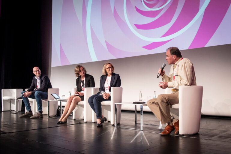
Despite research in the field is gaining momentum, real-world implementation of AI solutions remains complex, as highlighted by international experts’ panel discussions during an Education Session at the ESMO Breast Cancer 2025 in May (Berlin, Germany).
A co-pilot in digital pathology
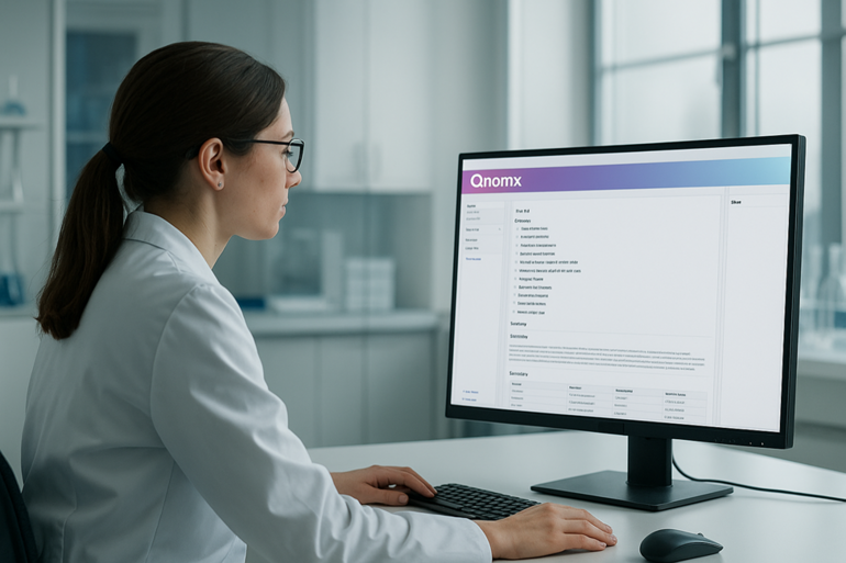
AI-guided pathology depends on converting traditional glass tumour slides into digital images via high-resolution scanners. While technology is expected to streamline laboratory workflows, several technical barriers, including data availability and quality, biases in model development and explainability, and the lack of adequate infrastructure are still limiting an AI-driven transformation of the field as reported in a systematic review conducted by representatives from the ESMO Precision Oncology Working Group (POWG), together with other international experts (Article in press, April 29, 2025).
According to Prof. Anne Vincent-Salomon of Institut Curie in Paris, France, who joined the expert panel, these issues risk to make some daily tasks more time-consuming rather than speeding them up. “For example, DICOM (Digital Imaging and Communications in Medicine) is not a shared standard yet, and different scanners used in different centres are associated to different whole slide image formats,” she noted during the session. “Also, we need that image management systems are completely interconnected with the laboratory management system, as well as a lot of storage capacity at our facilities.” She also added that beyond tools’ robustness that need to be shown in validation studies, practical usability is a driver factor to their implementation: “The ergonomy to use different tools in daily practice matters as it takes time to switch from one tool to another. And that is where access to AI through the existing PACS system is critical.”
Capturing cancer heterogeneity
Precision oncology is where AI solutions may bring novel opportunities to capture information on breast cancer that are not yet visible to expert human eyes about, including intratumour spatial variability, subclonality and tumour microenvironment. “AI brings a great advantage, and it is indeed possible to build robust models on a mixture of both data from different scanners and different labs,” said Dr Mattias Rantalainen, Karolinska Institute, Stockholm, Sweden, at the congress. With his research group, he developed Stratipath Breast, a CE-IVD marked AI solution for risk profiling of breast cancer patients which provides a more affordable alternative to molecular assays (Ann Oncol. 2022 Jan;33(1):89-98). “It integrates with some of the most common image management systems in the pathology lab, so there is no need to have a fully digital lab to its implementation,” he highlighted.
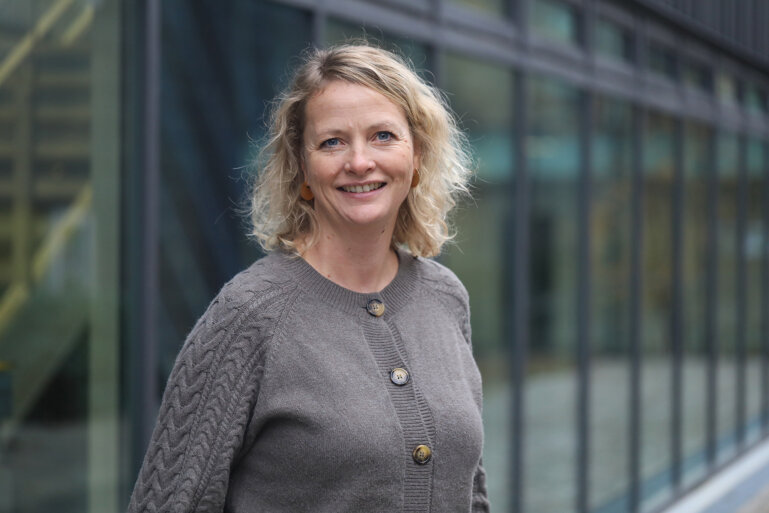
Rantalainen noted that there has been a peak in publications on foundation models last year, however “they are mostly trained on pan-cancer or pan-tissue data, which may be great for diversity, but not for capturing the more fine-grained information within each tissue type and each diagnosis.” Dr Anniina Färkkilä, University of Helsinki, Finland, agreed that the future of AI-driven oncology lies in training new foundation models with spatial biology and assay data merged with more simple layers like H&E.
“Tumour microenvironment is really a complicated, but coordinated and dynamic ecosystem,” she explained during her presentation. “There has been a huge surge of different spatial technologies emerging over recent years, including spatial proteomics assays, spatial transcriptomic assays, spatial metabolomics or lipidomics assays, producing huge amounts of data which are very complicated to analyze, so they are not scalable to clinics. AI tools can really help us to understand the tumour microenvironment.”
The bottleneck of cost and reimbursement
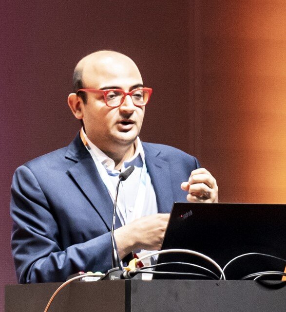
For Prof. Paul van Diest, University Medical Center Utrecht, The Netherlands, the pathology community shows a high interest to adapt to an emerging AI-guided scenario and willingness to integrate novel algorithms into daily practice. However, financial resources add to other technical issues currently limiting AI’s integration in clinical practice. “There is no reimbursement for AI tools, and this is a problem as AI costs. So, we need to find our own business cases for now by saving money elsewhere,” he explained in Berlin. A University Medical Center Utrecht’s experience showed that real-time clinical implementation of an AI-assisted workflow for detecting breast cancer metastases in sentinel lymph nodes (SNs) led to a lower use of immunohistochemistry per detected case, “with a cost saving of approximately 40,000 € annually” (Nat Cancer. 2024 Aug;5(8):1195-1205). But implementation is never straightforward, clarified van Diest who calculated that the process required the equivalent of “six man’s years invested” at the Dutch institute, so it may not be sustainable for all laboratories.
At the end of the session, he concluded: “Pathology is really cheap, it takes a tiny part in the global healthcare budget, so we as pathologists are entitled to some reimbursement to make sure we can make a real impact for our patients.”
AI & Digital Oncology: Resources in one place
Looking for further insights into how artificial intelligence and digital tools are impacting oncology? The ESMO AI & Digital Oncology Hub brings together expert perspectives, research updates, and thought leadership from across oncology.
It is a space where you can stay informed, discover resources, and follow the conversation on digital innovation in cancer research and treatment.
To further explore the transformative potential of AI in oncology, the very first ESMO AI and Digital Oncology Congress 2025, taking place from 12 to 14 November, will provide a dedicated platform focused on the latest advances in AI and digital technologies in cancer care.


