Pathology is going digital. More than 50 AIs cleared for diagnostic use are now available. This article summarises the status quo and gives an outlook of upcoming emerging technologies.
Authors:
- Volker Bruns, group manager Medical Image Analysis, Fraunhofer IIS, Erlangen, Germany
- Michaela Benz, chief scientist Digital Health & Analytics, Fraunhofer IIS, Erlangen, Germany
The content of this article was provided by Fraunhofer IIS and reflects the views of the authors / company. ESMO does not endorse the content of this publication and does not take any responsibility for the accuracy, completeness or reliability of the information contained in such article.
Digital pathology has firmly established itself, even in Germany. University hospitals have utilised scanners for years in research settings, where digitisation is essential for computational pathology, facilitating quantitative analysis and simplifying biomarker discovery. Recently, pathology institutes have also begun transitioning their clinical routine work to a digital workflow. These are large-scale undertakings unique to each university hospital, involving careful planning, securing budgets - such as through the "Krankenhauszukunftsgesetz" (Hospital Future Act) - engaging stakeholders, drafting tenders, and ultimately optimising the new processes.
Image management systems
Choosing a scanner from the myriad of available vendors is just one small facet of this transition. Hospitals must also decide whether to archive data on-premise or in the cloud. Additionally, selecting an Image Management System (IMS) is crucial; it serves as the “digital pathologist’s cockpit,” managing slide archival and functioning as an interface for AI tools from third-party vendors.
Some institutions opt to replace their lab information systems (LIS) as part of these digitisation initiatives. The lines between IMS and LIS are starting to blur, and future solutions may integrate both functionalities. When IMS and LIS remain separate, users open each system on different monitors, allowing them to communicate bidirectionally and ensuring the same case is accessible in both platforms. As with scanners, IMSes are available from a multitude of vendors (1). Many of them are radiology PACS vendors that are expanding their services into pathology - a field where the market is still in its early stages of development.
We anticipate that non-university hospitals and pathology practices will soon follow in a second wave of digitisation. A major motivating factor is the ability to work remotely, enabling pathologists to diagnose cases from home or during conferences. This flexibility enhances employer appeal, which is vital given the shortage of young pathologists. Diagnosing on computer screens is generally more ergonomic and less taxing on the eyes compared to traditional methods, where physical glass slides are viewed through a microscope while managing the LIS on a separate screen.
AI-assisted computational pathology
Moreover, digital pathology paves the way for AI-assisted computational pathology. The literature features thousands of publications demonstrating use of AI for interpreting histology scans. In the past few years, we've seen the introduction of the first AIs granted clearance for primary diagnostic use, obtaining CE-IVD or CE-IVDR marks. By the summer of 2025, approximately 50 such AI systems are available (2). Most of these AIs automate the evaluation of scores or tasks currently performed manually - such as assessing Ki67 proliferation or HER2 expression in breast cancer, PD-L1 expression in lung cancer, Gleason grading in prostate cancer, or detecting (micro)metastases in lymph node biopsies. The business case remains challenging though, as the extra costs for digitisation and AI are not reimbursed. An evolving category of AIs extends beyond mere automation to directly predict biomarkers or genetic mutations from H&E stains, eliminating the need for additional tests such as NGS. Here, AI may help cut costs, reduce turn-around times, and provide helpful guidance when the oncologist needs to select a therapy right away in urgent cases.
Given that histological scores often serve as proxies indicative of critical clinical outcomes such as tumor subtype, aggressiveness, or metastasis risk, an emerging category of AI products specialise in directly forecasting these endpoints. After all, conventional scores designed to be carried out manually need to be as simple as possible; AI can redefine these limitations. A first example of yet another new category of AI-only companion diagnostic tests was recently released. It quantifies TROP2 expression in the cell membrane and was shown to predict treatment response to a new ADC, while conventional immunohistochemistry did not (3). With the advent of ADCs, more such AI companion diagnostics may arrive in the near future.
Outlook: Spatial biology
To give an outlook on the technological side, spatial biology methods are revolutionising biomedical research and may soon find their way into the routine. While spatial transcriptomics, spatial proteomics or mass spectrometry imaging are still expensive and slow today, they paint a much fuller picture (both literally and figuratively). This holds even more true when combined in a multi-omics approach and provides a basis to derive much more accurate biomarkers. Another scenario is when biopsies are very hard to obtain, and only little tissue is available for selecting the best therapy. The future will tell if and where spatial biology will enter the clinical routine.
(1) Bruns, V. et al, Fraunhofer IIS, “The Digital Pathologist’s Cockpit: Image Management Systems (IMS) – Free Download of List of IMSes“, online, 13 August 2025: https://websites.fraunhofer.de/smart-sensing-insights/ims-vendors
(2) Bruns, V. et al, Fraunhofer IIS, “Digital Pathology AI Companies — List of Commercial AIs (CE-IVDR, FDA, RUO) “, online, 13 August 2025: https://websites.fraunhofer.de/smart-sensing-insights/digital-pathology-ais
(3) AstraZeneca, September 2024, online: https://www.astrazeneca.com/media-centre/press-releases/2024/novel-computational-pathology-based-trop2-biomarker-for-dato-dxd-was-predictive-of-clinical-outcomes-in-patients-with-nsclc-in-tropion-lung01-phase-iii-trial.html
(4) Fraunhofer IIS 2025, MIKAIA® studio Version 2.3 [Computer software]. RRID:SCR_025081. www.mikaia.ai
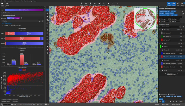
MIKAIA® is a digital pathology and spatial biology analysis software for life science researchers, developed by Fraunhofer IIS and distributed by partners worldwide (4). It comprises a multitude of algorithms & AIs usable off-the-shelf without programming or scripting expertise. Additional AIs can be interactively trained with the built-in AI author or integrated via the Python/R plugin API.
MIKAIA® lite freely available for download: www.mikaia.ai
Contact: volker.bruns@iis.fraunhofer.de