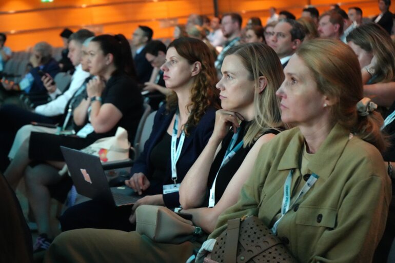
Experts' discussions during the ESMO Gastrointestinal Cancers Congress 2026 explored how AI is moving beyond image recognition to support surgery, pathology and image-guided intervention.
The value of AI in gastrointestinal (GI) oncology is becoming increasingly clear, as demonstrated by recent advances. In colorectal cancer, the MSIntuit algorithm detected microsatellite instability directly from routine haematoxylin and eosin (H&E) slides with 96–98% sensitivity (Nat Commun. 2023;14:6695), while deep learning models have been developed to predict lymph node metastasis from pre-treatment CT imaging in gastric cancer (eClinicalMedicine. 2024;75:102805).
Experts speaking at the ESMO Gastrointestinal Cancers Congress 2026 suggested the field is entering a new phase, where clinical value is proving to be just as important as technical performance.
Surgical AI looks beyond anatomy
According to Dr Marie Hanaoka, Institute of Science Tokyo, Japan, the field is moving towards "outcome-linked intelligence," in which AI-derived measurements made during surgery are linked to clinically meaningful patient outcomes rather than simply recognising anatomy or surgical workflow.
Presenting proof-of-concept data using the AI platform EUREKA X, Hanaoka quantified the preservation of loose connective tissue during robotic rectal cancer surgery. In a matched case-control study of 44 patients, AI-derived measures of tissue preservation were associated with postoperative urinary dysfunction. When combined with patient age, the model achieved an area under the curve of approximately 0.81.
Hanaoka suggested that future AI systems could provide real-time feedback during surgery. "Surgical AI is no longer just about recognition accuracy," she said. "We must ask whether this AI information is meaningful for our patients." Looking ahead, Hanaoka concluded: "The future is not AI versus surgeons. It is AI-enhanced surgeons."
Seeing what CT can’t
A similar evolution is taking place in image-guided intervention. Prof. Max Seidensticker, LMU Klinikum, Munich, Germany, described how MRI-guided interstitial brachytherapy is expanding treatment options for liver tumours that are unsuitable for thermal ablation. MRI guidance enables clinicians to visualise lesions that are often invisible on CT. Quoting Muhammad Ali, he said, "Your hands can't hit what your eyes don't see," highlighting the importance of accurate visualisation for precise catheter placement.
In the prospective MR BRIGHT study (Z Gastroenterol 2025; 63(01): e45), MRI-guided brachytherapy achieved significantly higher local recurrence-free survival than CT-guided treatment (95.1% versus 79.9%; p=0.027), while substantially reducing low-dose radiation exposure to healthy liver tissue (15% versus 43%). Looking ahead, Seidensticker described how machine learning algorithms could accelerate MRI image reconstruction and reduce artefacts, making real-time MRI guidance increasingly practical during intervention.
From promising algorithms to clinical tools
Pathology may be where AI's transition from research to routine clinical practice is most evident.
Prof. Magali Svrcek, Sorbonne Université, Paris, France, described AI as the product of four converging developments: an expanding biomarker landscape, increasing pathology workloads, the widespread adoption of digital pathology and persistent diagnostic variability. Together, these developments have transformed the pathology slide from something viewed under a microscope into "a source of quantitative biological data."
Presenting the ESMO basic requirements for AI-based biomarkers in oncology (EBAI) (Ann Oncol. 2026;37(3):414–430), Svrcek explained how algorithms are progressing from standardising existing biomarkers to predicting molecular alterations directly from H&E slides and ultimately discovering entirely new biomarkers. She also highlighted emerging models capable of recognising when they should not make a prediction (Ann Oncol. 2026;37(7):974-985).
For Svrcek, "The real challenge is not to demonstrate the technical performance or robustness of the algorithm, but to demonstrate the clinical value." Looking ahead, she concluded: "The future of pathology is not the replacement of pathologists by AI. It is the transformation of pathologists into experts in complex biological data, serving precision medicine and patient care."